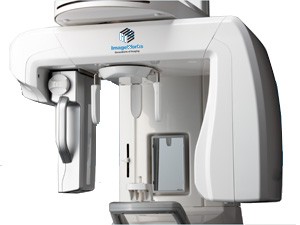
Panoura 18S Panoramic X-Rays The Panoura 18S is the most practical advanced 2D and 3D dental imaging system. Ideal for doctors who need Panoramic, Ceph and 3D capabilities all in one compact, low-cost platform.
DIRECT CONVERSION SENSOR
Most Panoramic CMOS sensors use lower cost, more generally available materials that are less sensitive to x-ray. Therefore, these sensors must convert x-ray to light before converting to a digital signal. This can result in image blurriness because the extra conversion step can cause the radiation to “fan out” and inadvertently trigger surrounding pixels. The Panoura has a unique sensor design that converts x-ray directly to digital, which contributes to a much sharper image.
Exquisite Images – Even With Suboptimal Positioning
Panoura 18S features auto-correction for suboptimal patient positioning. Without this, an already suboptimal image (from a pan with a lower-quality sensor) can be further degraded if the patient is not positioned optimally. With the Panoura, the very high data-capture sensor acquires many “layers” of image data. This allows the powerful software to auto-correct for suboptimal patient positioning. It’s worth noting that all of this data is available after the image is taken – so the image can be further optimized if desired. In the end, this assures exquisite images with the Panoura every time while being less dependent on operator training or the ability of the patient to remain stationary.
Easy 3D Upgrade
Our basic 2D panoramic unit is upgradeable to 3D, so there is peace of mind that the initial investment is protected if 3D capability is needed later on
Small Footprint
The Panoura’s small footprint makes it an incredibly practical solution for tight offices.
Flexible 3D FOV
Whether you need focused diagnosis and treatment, or an expanded view of the maxillary or mandibulars, the Panoura CBCT can dramatically improve the accuracy of an endodontic diagnosis or the implant treatment plan.
Powerful Implant Planning Tools
Everything you need for planning your implant surgery is included. Mark the mandibular canal and place implant fixtures from our library of real-size implant fixtures of all major manufacturers on the market today. From the simulation, you can decide the implant fixture size and drilling depth and direction
3D Zoom Accuracy
Our 3D rendering technology allows for detailed zoom-in capabilities without resolution loss. Instead of simply magnifying, 3D Zoom Cube re-renders the selected producing a high-quality, sharp image. Clearly visualize root canals and precisely map the alveolarnerve canal as you examine any cross section from all sides of the cube.
 The Panoura 18S is the most practical advanced 2D and 3D dental imaging system. Ideal for doctors who need Panoramic, Ceph and 3D capabilities all in one compact, low-cost platform.
DIRECT CONVERSION SENSOR
Most Panoramic CMOS sensors use lower cost, more generally available materials that are less sensitive to x-ray. Therefore, these sensors must convert x-ray to light before converting to a digital signal. This can result in image blurriness because the extra conversion step can cause the radiation to “fan out” and inadvertently trigger surrounding pixels. The Panoura has a unique sensor design that converts x-ray directly to digital, which contributes to a much sharper image.
Exquisite Images – Even With Suboptimal Positioning
Panoura 18S features auto-correction for suboptimal patient positioning. Without this, an already suboptimal image (from a pan with a lower-quality sensor) can be further degraded if the patient is not positioned optimally. With the Panoura, the very high data-capture sensor acquires many “layers” of image data. This allows the powerful software to auto-correct for suboptimal patient positioning. It’s worth noting that all of this data is available after the image is taken – so the image can be further optimized if desired. In the end, this assures exquisite images with the Panoura every time while being less dependent on operator training or the ability of the patient to remain stationary.
Easy 3D Upgrade
Our basic 2D panoramic unit is upgradeable to 3D, so there is peace of mind that the initial investment is protected if 3D capability is needed later on
Small Footprint
The Panoura’s small footprint makes it an incredibly practical solution for tight offices.
Flexible 3D FOV
Whether you need focused diagnosis and treatment, or an expanded view of the maxillary or mandibulars, the Panoura CBCT can dramatically improve the accuracy of an endodontic diagnosis or the implant treatment plan.
Powerful Implant Planning Tools
Everything you need for planning your implant surgery is included. Mark the mandibular canal and place implant fixtures from our library of real-size implant fixtures of all major manufacturers on the market today. From the simulation, you can decide the implant fixture size and drilling depth and direction
3D Zoom Accuracy
Our 3D rendering technology allows for detailed zoom-in capabilities without resolution loss. Instead of simply magnifying, 3D Zoom Cube re-renders the selected producing a high-quality, sharp image. Clearly visualize root canals and precisely map the alveolarnerve canal as you examine any cross section from all sides of the cube.
Manual Straight Arm
- Floor Mount 8’ Foot 9” ceiling
- H.T. Cables
- Manual Collimator
- DR adaptation for 17×17 and 14×17 application
- X-Ray Generator SHF 50KW, 3 phase 208 volt
- X-Ray Tube E7884X 150Kvp 300,000 HU 0, 6-1, 2mm
Generator
- SHF40, 40 kW, 500 mA HF-AP, 1-phase
Other generator sizes available
Tube
Toshiba, E7884FX, 300kHU tube, 150KVP, 0.6 x 1.2mm FS
Collimator
Manual collimator
Additional Accessories:
- Fixed enclosure trays for upright and table
- Custom rotating enclosure tray for tethered panel rotates for minimal handling for upright stands and tables
- Grids 8:1 and 10:1
- Weight bearing stand
- Weight bearing cap
- Grid cap
- Mobile table
- Generator integration supports most generators


