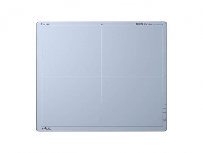
Overview
The Canon CXDI-701C Wireless Digital Radiography System is designed to help healthcare professionals attain a high level of performance, usability, and reliability. By utilizing technology and solutions that help accelerate exams and maintain productive workflow, practitioners can effectively accomplish more while providing quality care. In X-ray auto detection mode, this DR system can detect X-rays at exposure and shift to image acquisition mode automatically without the use of a typical generator interface. The CXDI-701C Wireless System builds upon the successful CXDI-70C Wireless DR system with its high-quality image sensors that provide high-resolution images at a low X-ray dose, and incorporates more customer-focused features, such as enhanced liquid intrusion protection, battery charging while in the detector with a wired option (sold separately), and impact detection. The CXDI 701C Wireless is light and easy to handle, weighing only 7.3 pounds.
Features
- Durable Cassette-size Detector:
The CXDI-701C Wireless Digital Radiography System is lightweight at 7.3 pounds (with the battery). It has the same dimensions as a standard ISO 4090 film cassette and fits into existing Bucky trays. For non-Bucky exposures, a handle unit (sold separately) is available for easy transport and attaching a grid. - High Resolution and High Sensitivity:
With a 125 micron pixel pitch, the CXDI-701C Wireless Digital Radiography System delivers exceptional spatial resolution. The high signal-to-noise performance of the Cesium Iodide (CsI) scintillator helps reduce the X-ray exposure to the patient. Tomography and other exams that require an exposure time of up to 3 seconds can be performed. - Wireless Range:
The CXDI-701C Wireless Digital Radiography System has a range that can be calibrated for signal strength. Depending upon the room in which the CXDI-701C Wireless DR System is used, the power can be managed and potential interference can be reduced. Should the wireless connection be lost, the unit will automatically attempt to reconnect and to prevent image loss. Acquired images are stored locally in the on-board memory. Once reconnected, the images can be retrieved. An optional wiring unit allows images to be transferred from a panel mounted in a Bucky to the control computer over hard wire. - 5 GHz for Fast Data Transfer:
The CXDI-701C Wireless DR Detector runs at 5 GHz and 2.4 GHz on an 802.11n network that provides signal clarity – resulting in fast data transfer speed. - Immediate Results:
An on-screen preview image is available approximately 1-2 seconds after exposure. An additional monitor (not included) can be added for the immediate viewing of high-resolution images in the emergency department or radiography suite. - Battery:
The battery lasts up to 6.5 hours and takes less than 3 hours to charge. The CXDI-701C Wireless DR System provides battery charging while in the detection with a wired option (sold seperately). An accessory battery charger (sold separately) can additionally charge up to 2 batteries at a time so a fresh battery can be continuously available. The control console includes a battery indicator for image count and power level monitoring. Additional batteries (sold separately) are also available. - Portable and Flexible:
Each individual CXDI-701C Wireless Digital Radiography System can be used in multiple X-ray rooms. Up to 10 detectors can be registered for use in any room. A check-in unit in each room identifies the particular detector being used there.
CXDI Control Software NE:
The CXDI-701C Wireless Digital Radiography System uses the Canon CXDI Control Software NE. The intuitive operation of this software gives a wide range of selections directly from the main menu, and images can be taken with three touches. The CXDI Control Software NE also has advanced image processing that shows the subtle details of trabecular bone structure and soft tissue in the same image. For more information, visit our CXDI Control Software NE webpage.
Detector | ||
| Method | Cassette Size Detector, Scintillator & Amorphous Silicon (a-Si) |
| Scintillator | Csl (Csl: TI) |
| Sensor | LANMIT (Large Area New-MIS sensor and TFT) |
| Pixel Pitch | 125 Microns |
| Pixels | 2,800 x 3,408 Pixels (9.5 Megapixels) |
| Imaging Area | 14 x 17 in (35 x 43 cm) |
| Standard Configuration | Sensor Unit, X-Ray Interface Box, Battery Charger, Detector Check-In Unit, Wireless Access Point (Including A/C Power) |
| ||
Battery | ||
| Life | Up to 6.5 Hours or 140 images |
| Recharge Time | Less Than 3 Hours |
| Performance | Approximately 140 Images |
| ||
Image Acquisition and Processing | ||
| Image Processing | Standard Advanced Multi-Frequency Processing |
| A/D | 16-bit |
| Grayscale Output | 12 bit or 16 bit selectable |
| Preview Image | Approximately 1-2 Seconds After X-ray Exposure |
| Cycle Time | Approximately 5-6 Seconds After X-ray Exposure |
| ||
Data Output and Network Connection | ||
| Wireless Standard | IEEE 802.11n (5 & 2.4 GHz) |
| DICOM | DICOM 3.0 Compatible, Print Management (SCU), Storage (SCU), MWL (SCU), MPPS (SCU) |
| ||
Electrical and Environmental | ||
| Operating Environment | Sensor Unit: 41-95°F (5-35°C), Humidity 30-80% RH (non-condensing) |
| ||
Physical Characteristics | ||
| Dimensions (W x L x H) | 15.1 x 18.1 x 0.6 in (384 x 460 x 15 mm) |
| Weight | 7.3 lbs (3.3 kg) (with battery) |
|
|
|
Specifications are subject to change without notice. | ||


